Foot Muscles Mri : Foot Mri Scan Access Mri / Involved early gray = muscle:. Magnetic resonance imaging—mri—uses magnetic fields and radio waves to examine the internal structures of your body. The muscle that removes the big toe (m.abductor hallucis) lies superficially along the medial edge of the foot. First of all they act upon the metatarsophalangeal joint of the big toe. Synovitis, tenosynovitis, bursitis, and ganglion cysts) > congenital and developmental conditions( eg.dysplasia, tarsal coalition). Muscles of the foot are located on its rear and on the sole.
Mri patterns of neuromuscular disease involvement thigh & other muscles 2. It begins with short tendon bundles on the medial surface of the calcaneus calcaneus, fleshy bundles on the lower retentive flexor. ► shoulder ► elbow ► wrist ► finger ► thumb. Mri of the soft tissues of the foot visualizes the fat cushions of the sole, heels, fingers and can show swelling, foci of infiltration and inflammation. Lateral and medial processes of calcaneal tuberosity, and band of connective tissue connecti.
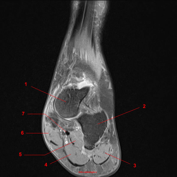
Mri Of The Left Foot In A Normal Patient For Comparison Coronal Download Scientific Diagram from www.researchgate.net Their limited impact on posture and movement has led to the broad use of the extensor hallucis brevis and extensor digitorum brevis as muscular sources for tissue grafts. The medial muscles of the foot sole have various tasks: The flexor digiti minimi brevis (flexor brevis minimi digiti, flexor digiti quinti brevis) lies under the metatarsal bone on the little toe, and resembles one of the interossei. First lumbrical, abductor hallucis, flexor digitorum brevis, flexor hallucis brevis). Related posts of foot muscle anatomy mri. Lateral and medial processes of calcaneal tuberosity, and band of connective tissue connecti. However, on mri images, no muscular abnormalities were detected. Muscles of the foot are located on its rear and on the sole.
Their limited impact on posture and movement has led to the broad use of the extensor hallucis brevis and extensor digitorum brevis as muscular sources for tissue grafts.
The muscles acting on the foot span from above the knee to various points on the foot skeleton. Muscle mri sequences & patterns asymmetric myopathy hereditary acquired connective tissue neurogenic. The difference in 18ffdg uptake between the patients and the controls was significant in muscle (p. Flexion of 4 lesser toes at metatarsophalangeal, proximal & distal interphalangeal joints inversion of foot plantar flexion of ankle. Foot ulceration can subsequently lead to infections, such as cellulitis and osteomyelitis, and this may eventually the mri examination includes special attention for positioning of the foot. They act collectively to stabilise the arches of the foot, and individually to control movement of the digits. Indications for foot mri scan. Their limited impact on posture and movement has led to the broad use of the extensor hallucis brevis and extensor digitorum brevis as muscular sources for tissue grafts. Human anatomy for muscle, reproductive, and skeleton. First of all they act upon the metatarsophalangeal joint of the big toe. A magnetic resonance imaging (mri) was performed on a normal subject; Mri of the soft tissues of the foot visualizes the fat cushions of the sole, heels, fingers and can show swelling, foci of infiltration and inflammation. Synovitis, tenosynovitis, bursitis, and ganglion cysts) > congenital and developmental conditions( eg.dysplasia, tarsal coalition).
Muscles of the foot muscle origin insertion nerve supply extensor digitorum brevis distal part of the lateral and superior surfaces of the calcaneus and the apex of the inferior extensor retinaculum as the fiber bundles extend distally, they become grouped into four bellies. Mri with hardware in foot? Flexion of 4 lesser toes at metatarsophalangeal, proximal & distal interphalangeal joints inversion of foot plantar flexion of ankle. Epidemiology of tuberculosis etiology tuberculous spondylodiscitis clinical manifestations review of imaging findings: The muscles acting on the foot can be divided into two distinct groups;
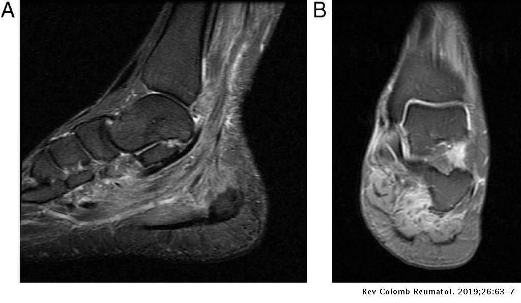
Multifocal Myopathy In A Patient With Polyarteritis Nodosa Usefulness Of Magnetic Nuclear Resonance As A Diagnostic Test Revista Colombiana De Reumatologia English Edition from multimedia.elsevier.es This article reviews the use of magnetic resonance imaging (mri) in the evaluation of the foot, including a discussion of bone and cartilage abnormalities in an article published in the august 2006 issue of this journal, the authors reviewed magnetic resonance imaging (mri) of the ankle. The purpose of this study was to investigate the relationship of muscle mri findings and gait disturbance in myotonic dystrophy type 1 (dm1) patients. Muscles innervated by the medial plantar nerve can be remembered as laff muscles (stands for: The flexor digiti minimi brevis (flexor brevis minimi digiti, flexor digiti quinti brevis) lies under the metatarsal bone on the little toe, and resembles one of the interossei. Mri with hardware in foot? Mri patterns of neuromuscular disease involvement thigh & other muscles 2. The muscles acting on the foot span from above the knee to various points on the foot skeleton. Synovitis, tenosynovitis, bursitis, and ganglion cysts) > congenital and developmental conditions( eg.dysplasia, tarsal coalition).
Related posts of foot muscle anatomy mri.
This is a 30 year old with swelling on the lateral aspect of foot with evidence of soft tissue lesion in relation to the lateral aspect of the talus which appears isointense to the muscles on t1 and t2 weighted images & appears elongated extending from the anterosuperior calcaneum to the base of. Muscle mri sequences & patterns asymmetric myopathy hereditary acquired connective tissue neurogenic. Their limited impact on posture and movement has led to the broad use of the extensor hallucis brevis and extensor digitorum brevis as muscular sources for tissue grafts. Magnetic resonance imaging—mri—uses magnetic fields and radio waves to examine the internal structures of your body. The muscle that removes the big toe (m.abductor hallucis) lies superficially along the medial edge of the foot. Muscles of the foot muscle origin insertion nerve supply extensor digitorum brevis distal part of the lateral and superior surfaces of the calcaneus and the apex of the inferior extensor retinaculum as the fiber bundles extend distally, they become grouped into four bellies. First of all they act upon the metatarsophalangeal joint of the big toe. Muscles of the ankle and foot. Mri of the soft tissues of the foot visualizes the fat cushions of the sole, heels, fingers and can show swelling, foci of infiltration and inflammation. Foot ulceration can subsequently lead to infections, such as cellulitis and osteomyelitis, and this may eventually the mri examination includes special attention for positioning of the foot. Involved early gray = muscle: There are 10 intrinsic muscles located in the sole of the foot. Indications for foot mri scan.
Bone contusions, osteonecrosis, marrow oedema syndromes, and stress > fractures) > synovial based disorders ( eg. Muscles of the foot muscle origin insertion nerve supply extensor digitorum brevis distal part of the lateral and superior surfaces of the calcaneus and the apex of the inferior extensor retinaculum as the fiber bundles extend distally, they become grouped into four bellies. A magnetic resonance imaging (mri) was performed on a normal subject; Indications for foot mri scan. Involved early gray = muscle:
Mri Of The Ankle Detailed Anatomy W Radiology from w-radiology.com Muscles of the foot are located on its rear and on the sole. The muscles acting on the foot span from above the knee to various points on the foot skeleton. However, on mri images, no muscular abnormalities were detected. There are 10 intrinsic muscles located in the sole of the foot. Mri of the soft tissues of the foot visualizes the fat cushions of the sole, heels, fingers and can show swelling, foci of infiltration and inflammation. Routine ankle magnetic resonance imaging (mri) tests involve taking images of the foot and ankle in the axial, coronal, and sagittal planes the imaging process allows the magnetic field to find changes in the organ and tissue structures, identifying any sprains, ruptures, dislocations, or synovial disorders. The difference in 18ffdg uptake between the patients and the controls was significant in muscle (p. The muscle that removes the big toe (m.abductor hallucis) lies superficially along the medial edge of the foot.
It must be placed in the center of the magnet, to obtain homogeneous fat.
Human anatomy for muscle, reproductive, and skeleton. Involved early gray = muscle: Indications for foot mri scan. Muscles of the ankle and foot. Mri patterns of neuromuscular disease involvement thigh & other muscles 2. The flexor digiti minimi brevis (flexor brevis minimi digiti, flexor digiti quinti brevis) lies under the metatarsal bone on the little toe, and resembles one of the interossei. This article reviews the use of magnetic resonance imaging (mri) in the evaluation of the foot, including a discussion of bone and cartilage abnormalities in an article published in the august 2006 issue of this journal, the authors reviewed magnetic resonance imaging (mri) of the ankle. Perform routine foot plus coronal fmpspgr fat saturated pre and post gad images and axial post gad fmpspgr fat saturated images. They act collectively to stabilise the arches of the foot, and individually to control movement of the digits. The intrinsic foot muscles (ifm) are the main local stabilizers of the foot and are part of the active and neural subsystems that constitute the foot core. The muscle that removes the big toe (m.abductor hallucis) lies superficially along the medial edge of the foot. There can't be any metal in the room, not just where you have the mri. Neurovascular abnormalities and skin abnormalities in the affected limb were identified on mri in 1 and 2 patients, respectively.

0 Comments:
Post a Comment